




Monday, 20 July 2026 01:30 PM
Product Announcements
The Falcon-821CRS is an 8MP USB Camera built on the Onsemi AR0821 HyperLux sensor and positioned as an HDR ophthalmic camera for anterior segment imaging, corneal examination, and iris imaging under variable illumination conditions. The camera delivers 4K LI-HDR output with on-chip auto exposure, full UVC compliance, and a compact 38mm form factor for slit lamp attachment and ophthalmic instrument integration. It requires no driver installation on Windows, Linux, or Android and connects to any clinical workstation over a single USB 3.2 cable.
FORT WORTH, TX / ACCESS Newswire / July 20, 2026 / Vadzo Imaging, a provider of embedded vision camera products, today positions the Falcon-821CRS as an AR0821 Anterior Segment Camera for ophthalmic clinical and OEM imaging applications. The Falcon-821CRS operates as an HDR Ophthalmic Camera delivering 8MP 4K output from the Onsemi AR0821 HyperLux sensor with on-chip LI-HDR capable of up to 120 dB dynamic range. As a 4K HDR USB Camera, the module integrates into slit lamp attachments, anterior segment imaging systems, and ophthalmic diagnostic workstations via USB 3.2 with no driver installation required. Vadzo engineered this camera to address the specific imaging challenges of the anterior eye: the extreme contrast between a focused slit lamp beam and the surrounding ocular structures, the specular reflections from corneal surfaces, and the fine spatial detail required for iris pattern analysis and corneal feature documentation.
Sensor and Camera Overview
The Falcon-821CRS is an AR0821 Corneal Imaging Camera and AR0821 Slit Lamp Camera built on the Onsemi AR0821 HyperLux CMOS sensor and paired with a high-performance ISP. The AR0821 is an 8MP (3848 x 2168) color rolling shutter sensor with a 1/1.7-inch optical format and 2.1 µm pixel size. The sensor integrates on-chip LI-HDR capable of up to 120 dB dynamic range, which is the specific performance characteristic required to capture detail across the slit lamp's bright illuminated optical section and the adjacent dark ocular tissue simultaneously. The AR0821 sensor's color fidelity, full well capacity per pixel, and low read noise make it a Wide Dynamic Range Ophthalmic Camera suited for imaging the specular reflecting corneal surface, the pigmented iris stroma, and the limbal vasculature within a single calibrated exposure. This combination of 4K spatial resolution and 120 dB dynamic range establishes the Falcon-821CRS as a 4K HDR USB Camera and High Fidelity Eye Camera for anterior segment imaging.
The Falcon-821CRS is a compact 8MP USB Camera with a USB 3.2 interface housing the Onsemi AR0821 sensor, ISP, S-Mount (M12) lens assembly, and UVC-compliant connectivity for ophthalmic imaging. The S-Mount lens holder accommodates standard M12 optics with magnifications calibrated for slit lamp coupler distances and direct anterior segment working distances. Auto exposure operates continuously in hardware without host processor load, compensating for slit width changes during examination without clinician intervention. The camera enumerates as a standard UVC device on Windows, Linux, and Android without driver installation and draws all operating power from the USB host port at 5V. Output modes include 4K, 1080p, 720p, and VGA, supporting full resolutions, still capture, and compressed live preview in ophthalmic workstation software. As an 8MP USB Camera, the Falcon-821CRS captures native 3848 × 2168 images, enabling detailed documentation of corneal structures, iris anatomy, and anterior segment features. The module is designed as an 4K HDR USB Camera for seamless integration into slit lamps, ophthalmic diagnostic systems, and OEM medical imaging devices requiring plug-and-play USB connectivity. For more insights on high-resolution USB cameras for medical devices: High Resolution USB Camera for Medical Devices | Vadzo Imaging
Key specs: 8MP (3848 x 2168) | Onsemi AR0821 HyperLux 1/1.7-inch 2.1 µm pixel | Color | Rolling Shutter | Fixed Focus | Auto Exposure and LI-HDR | USB 3.2 Gen1 | UVC Compliant | 4K / 1080p / 720p / VGA | S-Mount (M12) | Windows / Linux / Android | Compact 38mm Module
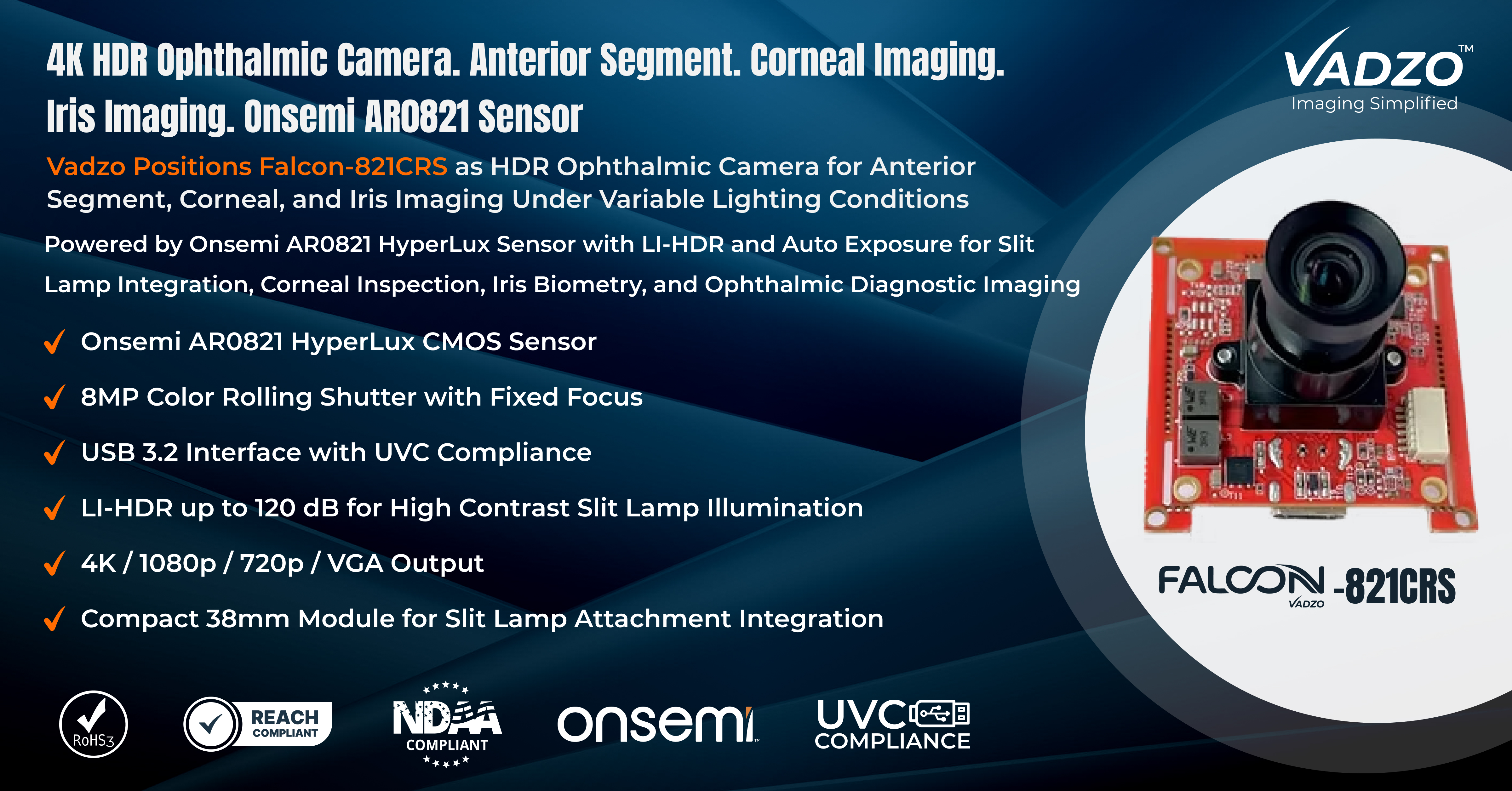
Key Capabilities of the Onsemi AR0821 4K HDR Ophthalmic Camera
On-chip LI-HDR for Slit Lamp Illumination Contrast: A slit lamp projects a narrow, collimated beam of light at high luminance through the cornea, lens, or anterior chamber. The illuminated optical section is many orders of magnitude brighter than the surrounding ocular tissue in diffuse illumination. A standard camera exposed for the bright slit section clips the tissue immediately outside the beam, and a camera exposed for the background renders the slit section entirely saturated. The Onsemi AR0821 on-chip LI-HDR captures the sensor at multiple exposures within a single frame readout cycle and merges them at the sensor level, preserving detail across the full 120 dB range from the bright slit interior to the dark iris periphery. This makes the Falcon-821CRS a true 8MP Slit Lamp Camera that documents corneal stroma cross sections, endothelial reflections, and iris surface texture within the same captured frame. As a Slit Lamp Camera Module, its HDR resolves the slit beam illumination contrast without requiring the clinician to adjust camera exposure manually between narrow slit and broad diffuse modes. The 4K Slit Lamp Camera resolution ensures that fine corneal epithelial layers, Bowman's membrane, and stromal fiber patterns within the optical section are resolved at the pixel level rather than interpolated.
Color Fidelity for Iris and Corneal Tissue Discrimination: Iris stroma pigmentation, limbal stem cell zone coloration, corneal neovascularization, and conjunctival injection all carry diagnostic significance expressed as color differences that may be subtle under clinical illumination. A camera with poor color science shifts iris brown toward orange, renders corneal neovascularization as ambiguous pink rather than clearly vascular red, and mutes the yellow-green hue of lipid deposits in corneal arcus. The AR0821 Bayer filter array and ISP color matrix preserve the spectral separation between these tissue categories with the color accuracy required for photographic documentation and serial comparison. As an AR0821 Iris Imaging Camera, the Falcon-821CRS captures iris crypt geometry, collarette boundaries, and pupil margin irregularities with faithful pigmentation rendering. As a Corneal Imaging Camera, it documents neovascular vessel caliber, corneal haze boundaries, and epithelial staining patterns with the color discrimination that fluorescein and rose bengal-stained examinations require. Vadzo's ISP calibration for the Iris Imaging Camera application is tuned for slit lamp illumination color temperature rather than the daylight standard used for consumer camera tuning.
4K Spatial Resolution for Anterior Segment Structural Detail: Anterior segment structures, including the corneal endothelial mosaic, iris crypt network, limbal palisades of Vogt, and pupil margin irregularities, require sufficient spatial resolution to be documented with diagnostic accuracy at the working distances typical of slit lamp couplers and anterior segment camera attachments. At 1080p, magnified views of the corneal endothelium produce interpolated crops that cannot reliably count or classify cell morphology. At 4K, the Falcon-821CRS captures 8MP native sensor pixels across the same field of view, providing the spatial sampling needed for cell density estimation and morphological classification. As an 8MP Anterior Segment Camera, the sensor delivers 3848 x 2168 native resolution that resolves the fine trabecular structure of iris crypts, the precise geometry of pupil margin irregularities, and the spatial distribution of limbal stem cells. For the clinical Anterior Segment Camera application, this means that a single captured frame at full resolution contains sufficient pixel density for both wide field anatomical documentation and zoomed inspection of focal features without capturing a second image at higher magnification. The Anterior Eye Imaging Camera 4K output also supports digital zoom within ophthalmic workstation software without the loss of diagnostic detail that 1080p crops produce.
Automatic Exposure Control Across Changing Examination Illumination: Ophthalmic examinations with a slit lamp involve continuous adjustment of slit width, illumination intensity, and beam angle as the clinician surveys different anterior segment regions. Each adjustment changes the overall scene of luminance significantly. Manual camera exposure adjustment during examination disrupts the clinical workflow and introduces delays that prevent continuous video documentation of dynamic findings. The Falcon-821CRS on-chip auto exposure operates continuously in hardware, adjusting gain and integration time frame by frame in response to scene luminance changes without host processor commands. As an Auto Exposure Ophthalmic Camera, it maintains consistent image brightness as the clinician transitions from broad diffuse illumination to narrow slit section, from direct to retro illumination, and from low magnification overview to high magnification focal examination. The HDR Corneal Camera auto exposure operates within the LI-HDR capture pipeline, maintaining both the exposure balance between slit and background and the overall image brightness simultaneously. For an Ocular Surface Camera capturing the tear film, conjunctival surface, and limbal zone under variable illumination, this automatic control removes the exposure management burden from the examiner entirely.
UVC Compliance for Direct Integration with Ophthalmic Workstations: Ophthalmology clinics and hospital eye departments operate imaging workstations running electronic medical record software, and imaging capture applications on Windows platforms with strict IT security policies. A slit lamp camera requiring a proprietary capture driver competes with EMR software for system resources, requires IT authorization for installation, and creates version dependency issues across Windows updates. The Falcon-821CRS connects as a Slit Lamp USB Camera that enumerates via the USB Video Class standard natively supported in Windows without any vendor driver. As a UVC Ophthalmic Camera, it appears through the standard DirectShow and Media Foundation APIs, making it accessible to any imaging capture application, EMR photo documentation module, or telemedicine platform that reads from the OS camera API. Any USB Slit Lamp Camera that relies on a proprietary SDK becomes dependent on vendor software maintenance timelines. The Falcon-821CRS avoids that dependency entirely by using the OS native interface, ensuring that the camera remains functional across Windows version updates without requiring camera-specific software patches from Vadzo.
OEM Ready Module for Ophthalmic Instrument Integration: Ophthalmic instrument OEMs developing slit lamp cameras, anterior segment imaging systems, portable iris imagers, and corneal topography attachments need a camera core with documented mechanical dimensions, compliance certification, and engineering support throughout the device development cycle. The Falcon-821CRS is available as an OEM Anterior Segment Camera module with RoHS 3 and REACH compliance documentation, mechanical drawings with lens mount geometry, and USB descriptor specifications for device firmware customization. As an OEM Eye Camera Module, the S-Mount (M12) lens holder supports magnification selection for specific slit lamp coupler optical paths, anterior segment working distances of 50 mm to 150 mm, and macro configurations for corneal surface imaging. Vadzo provides Ophthalmology Camera Module engineering consultation, including ISP color calibration for slit lamp illumination spectra, custom focus distance setting, and USB descriptor tuning for certified medical device integration. The Onsemi AR0821 Ocular Camera core is production-proven across multiple Vadzo camera deployments and supported with direct engineering access throughout the OEM device qualification cycle.
"Anterior segment imaging places specific demands on a camera sensor that general-purpose industrial cameras do not address. You need 120 dB dynamic range to handle the slit beam contrast, you need color accuracy calibrated for slit lamp illumination rather than daylight, and you need 4K spatial resolution to document corneal endothelial morphology and iris crypt geometry with confidence. The AR0821 HyperLux sensor delivers all three in a 38mm USB module with no driver requirements and bus power over USB. Vadzo positions the Falcon-821CRS for ophthalmology because the sensor architecture genuinely fits the application physics, not because it is the nearest available camera. We tune the ISP for clinical ophthalmic color and validate the HDR response against real slit lamp illumination conditions so that the module arrives ready for integration rather than requiring the OEM to perform that application work." - Alwin Vincent, Product Manager, Vadzo Imaging.
Applications
Slit Lamp Anterior Segment Documentation: Clinical slit lamp examination requires photographic documentation of corneal pathology, anterior chamber findings, iris lesions, and lens changes for patient records and longitudinal comparison. The Falcon-821CRS operates as a Slit Lamp Imaging Camera attached to the slit lamp camera coupler via S-Mount (M12) optics calibrated for the coupler's image plane distance. As a Clinical Ophthalmic Camera, it captures full 8MP images and 4K video of slit section findings under both focused slit and broad diffuse illumination without requiring manual exposure adjustment between modes. The HDR output preserves detail across the full anterior segment field from the corneal surface to the anterior iris plane within a single capture.
Corneal Examination and Endothelial Imaging: Corneal examination applications, including specular microscopy attachment cameras, corneal topography systems, and direct slit lamp for corneal documentation, require a camera that resolves the fine cellular and structural detail of the corneal layers. The Falcon-821CRS functions as a Corneal Inspection Camera delivering 4K resolution output through S-Mount macro-optics calibrated for corneal working distances. The 2.1 µm pixel size and 8MP frame capture the spatial detail of corneal epithelial cell patterns, stromal nerve fiber bundles, and endothelial cell morphology at the imaging magnifications achievable with standard slit lamp coupler configurations. As a Cornea Examination Camera, the LI-HDR manages the specular reflection from the anterior corneal surface that otherwise overwhelms standard camera exposure and obscures the cellular detail immediately adjacent to the bright reflection zone.
Iris Imaging and Anterior Eye Diagnostics: Iris imaging applications, including iris biometry, pupil margin analysis, iris lesion documentation, and congenital anomaly recording, require a camera that captures the full chromatic and spatial complexity of the iris stroma with sufficient resolution for pattern-level analysis. The Falcon-821CRS serves as an Anterior Eye Diagnostic Camera delivering color accurate 4K images of iris surface architecture, including crypts, collarette, and radial fiber bundles under slit lamp diffuse illumination. As an Eye Examination Camera, the auto exposure and LI-HDR maintain consistent iris surface exposure across changes in pupil dilation state, which alter the proportion of iris surface visible and the reflectance balance between the iris and the darker pupil aperture.
Digital Ophthalmology and Remote Clinical Review: Digital ophthalmology programs collecting anterior segment images for remote specialist review, AI-assisted screening, and population-level ocular health surveys require a standardized camera module that delivers consistent image quality across multiple sites and operators. The Falcon-821CRS operates as a Medical Eye Imaging Camera with UVC enumeration behavior and ISP calibration that remains consistent across all units, enabling meaningful cross-site image comparison. As a Digital Ophthalmology Camera, the module integrates into portable screening systems used in school vision programs, rural eye camps, and occupational health screenings, where consistent documentation quality supports algorithmic screening and remote clinical review by ophthalmologists who were not present at the examination.
High Resolution Anterior Eye Imaging Systems: Anterior eye imaging systems requiring high spatial resolution for research applications, custom corneal topography attachments, and precision iris biometry devices benefit from the AR0821 sensor's 8MP native resolution and 2.1 µm pixel geometry. As an Ophthalmic Slit Lamp Camera System integrated through S-Mount optics with custom magnification, the Falcon-821CRS delivers more resolved pixels per degree of anterior eye anatomy than lower resolution camera modules. As a High Resolution Eye Camera, it captures iris crypt dimensions, limbal palisade spacing, and corneal epithelial cell diameter at the pixel level rather than through interpolated magnification, supporting both clinical research and quantitative anterior segment analysis workflows.
OEM Ophthalmic Device and Instrument Integration: Ophthalmic instrument manufacturers developing portable slit lamp camera systems, handheld anterior segment imagers, and clinical iris documentation devices need a camera core with production-ready compliance documentation and engineering support for device certification. The Falcon-821CRS integrates as an Eye Imaging Camera Module within custom ophthalmic instrument enclosures, with Vadzo providing mechanical drawings, USB descriptor documentation, and ISP calibration data for the specific optical path of the OEM device. As a USB Ophthalmology Camera, the module connects directly to the device's embedded host processor over USB 3.2 without requiring a dedicated camera interface board or proprietary firmware layer, simplifying the hardware design and reducing certification scope.
Frequently Asked Questions
Q: What makes on-chip HDR essential for slit lamp photography rather than software HDR processing?
A: Software HDR blends multiple images captured sequentially - a standard bright exposure and a dark exposure taken at different time points. For a static scene, this can work acceptably, but a slit lamp examination involves patient movement, eye microsaccades, and clinician-directed lamp position changes between frames. When the two sequential exposures are captured milliseconds apart, any patient's motion creates a misalignment artifact in the merged result that produces blurred edges and ghost structures at the corneal surface boundary and iris margin. On-chip LI-HDR in the AR0821 captures both the bright and dark exposures within a single sensor readout cycle using line interleaved exposure integration, so both exposure levels are sampled from the same moment in time. Patient micromotion between the short and long exposures is minimized to a fraction of a single frame period, making the merged HDR output artifact-free at the fine structural scales relevant to corneal and iris imaging. The practical result is that a slit lamp using Vadzo's camera captures a motion-free 120 dB HDR image in a single shutter actuation, which is what clinical workflow and precise ophthalmic documentation require.
Q: How does 4K resolution improve corneal endothelial cell documentation compared to standard 1080p cameras?
A: Corneal endothelial cells are approximately 20 µm in diameter and are imaged through a slit lamp at moderate magnification using specular reflection or endothelial contact microscopy techniques. At 1080p with a typical slit lamp coupler magnification, each cell occupies only a few pixels in the captured image, making morphological classification and density estimation unreliable. At 4K, the same optical magnification produces four times as many pixels across the same field of view, allowing each endothelial cell to be represented by substantially more pixels. This improved spatial sampling improves cell boundary delineation, enables more accurate automated cell counting algorithms, and allows the reviewing clinician to assess polymegethism and pleomorphism from the digital image rather than requiring repeat examination. Vadzo's 8MP native resolution output provides this spatial detail without digital upscaling, meaning every pixel in the 4K output corresponds to actual sensor data. The difference between native 4K and upscaled 4K is significant for quantitative corneal analysis: upscaled images interpolate detail that native resolution cameras resolve, and that interpolated detail degrades algorithm accuracy in automated endothelial cell density tools.
Q: How does Vadzo calibrate the color profile of its ophthalmic camera for slit lamp illumination?
A: Standard camera ISP color matrices are calibrated under D65 daylight illumination at 6500 K, which is the standard for consumer and general industrial cameras. Slit lamp illumination uses xenon arc or LED sources with color temperatures typically between 4500 K and 6000 K and spectral power distributions that differ significantly from D65 in their short wavelength blue content. When a camera calibrated for D65 is used under slit lamp illumination, the color matrix produces a slight warm shift in tissue rendering that alters the perceived color of corneal neovascularization, iris pigmentation gradients, and conjunctival injection. Vadzo calibrates each Falcon-821CRS ISP color matrix specifically against representative slit lamp illumination spectra, producing a color profile where the white balance, saturation balance, and hue rotation are matched to typical slit lamp light sources rather than to daylight. The result is that iris brown tones, corneal transparency, and vascular red channels are rendered with the color accuracy that clinical photographers and ophthalmic researchers expect from a dedicated ophthalmic imaging instrument. OEM customers integrating the module into instruments with custom optical filters or non-standard illumination sources can request a custom ISP profile matched to their specific spectral conditions.
Q: How does UVC compliance simplify integration with ophthalmology EMR systems and imaging software?
A: Ophthalmology EMR systems and clinical imaging platforms access camera hardware through the operating system's standard video capture API - DirectShow on Windows, V4L2 on Linux, and AV Foundation on macOS. Any camera that enumerates as a UVC device is accessible through these APIs without custom integration work, because the EMR vendor has already written camera access code against the standard OS API. This means that when the Falcon-821CRS connects to a clinical workstation, the EMR photo documentation module sees it immediately as a capture source alongside any previously configured camera. There is no SDK license, no capture DLL to deploy alongside the EMR application, and no integration testing needed by the EMR vendor. For a hospital IT department evaluating a new ophthalmic camera, UVC compliance means the camera passes through their standard device approval process as a USB webcam class device rather than as custom medical device software, significantly reducing the approval timeline. Vadzo provides USB descriptor documentation confirming exactly how the Falcon-821CRS enumerates under each supported OS, allowing IT departments and EMR integrators to verify compatibility before deployment without requiring a physical device evaluation unit.
Q: What engineering resources does Vadzo Imaging provide to support ophthalmic instrument OEM development projects?
A: Vadzo Imaging provides ophthalmic OEM development teams with a complete package beyond the camera module itself. The engagement begins with a lens selection consultation: Vadzo's applications team evaluates the OEM's slit lamp coupler geometry, working distance, and required field of view and recommends the S-Mount optic that delivers the correct magnification and depth of field at the target anterior segment working distance. For ISP color calibration, Vadzo provides a factory-calibrated default profile and can produce a custom profile matched to the OEM device's specific illumination source and optical filter stack. Mechanical integration is supported with dimensioned drawings of the module, lens holder geometry, and mounting interface specifications. USB descriptor documentation covers VID/PID configuration for device-specific enumeration, descriptor tables for all supported resolutions and frame rates, and UVC control layout for exposure, gain, and white balance. Compliance documentation for RoHS 3 and REACH is provided with each module and updated to current directive versions for regulatory submissions. Throughout the development and clinical validation phase, direct engineering support from Vadzo's product team is accessible without escalation through a general support queue. Vadzo's goal is to ensure that the ophthalmic instrument enters clinical validation with a fully characterized imaging core rather than requiring the OEM team to independently resolve camera performance issues during the final stages of device development.
Availability
HDR Ophthalmic Camera Modules based on the Onsemi AR0821 HyperLux sensor are available now for evaluation and production orders. Evaluation kits for the Falcon-821CRS include the camera module with S-Mount fixed focus lens, USB 3.2 cable, and integration documentation with no minimum order requirement. Browse the full Vadzo medical and ophthalmic imaging portfolio at https://www.vadzoimaging.com/ or contact Vadzo at [email protected] to request an evaluation kit or discuss ophthalmic OEM integration requirements. For more insights on high-resolution USB cameras for medical devices: High Resolution USB Camera for Medical Devices | Vadzo Imaging
About Vadzo Imaging
Vadzo Imaging is a global provider of embedded vision solutions and delivers high-performance camera technologies and imaging platforms for applications in robotics, industrial automation, UAVs, edge AI, and medical systems. Its products are designed for seamless integration with leading embedded platforms. Vadzo supports customers through hardware customization, firmware development, and module-level drivers, enabling faster development and deployment of vision-based systems.
Media Contact
Alwin Vincent
Vadzo Imaging
Email: [email protected]
LinkedIn: Vadzo Imaging
YouTube: Vadzo Imaging
X: Vadzo Imaging
SOURCE: Vadzo Imaging