
Monday, 22 June 2026 02:00 PM
Company Update
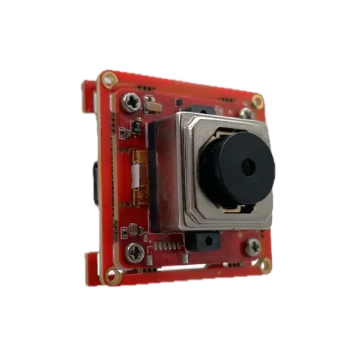
Vadzo Imaging's Falcon-521CRH is a 5MP color USB 3.2 Gen 1 camera module built on the Onsemi AR0521 sensor, delivering VCM autofocus, low-light performance, and UVC plug-and-play compliance for OEM integration into dermatology imaging systems, ENT examination platforms, teledermatology carts, wound documentation devices, and otoscope modules where accurate close-range tissue visualization at variable working distances is the primary engineering requirement.
FORT WORTH, TX / ACCESS Newswire / June 22, 2026 / Vadzo Imaging, a provider of embedded vision camera products, today introduces the Falcon-521CRH, a 5MP HDR USB 3.2 Gen 1 camera module built on the Onsemi AR0521 sensor and designed for OEM engineers developing dermatology imaging systems, ENT examination devices, wound documentation platforms, and teledermatology carts. Combining VCM autofocus with a 2592 x 1944 color sensor and USB 3.2 Gen 1 UVC compliance, the Falcon-521CRH connects directly to clinical workstations and embedded computing platforms without custom driver development, making it a production-ready module for embedded clinical device programs across the medical imaging market.
Imaging Challenges in Clinical Dermatology and Close-Range Tissue Examination
Imaging systems embedded in dermatology examination devices operate under constraints that consumer-grade USB camera products and fixed-focus board-level modules do not address. Skin lesion documentation requires sufficient spatial resolution to capture surface texture, color distribution, border irregularity, and morphological detail at working distances that vary between macro-close mucosal imaging and standard examination distance. ENT scope attachment modules and otoscope-integrated camera products must maintain sharp focus within the narrow working range of an ear canal or nasal passage examination, where the physician's hand position varies between patients and between examination passes. Wound imaging platforms used in post-operative care and chronic wound monitoring require accurate color rendition to track tissue tone, perfusion indicators, granulation development, and slough boundaries across sequential documentation sessions. Fixed-focus camera products fail across all of these scenarios because a single calibrated working distance cannot cover the range of examination geometries that a production clinical device must support.
Onsemi AR0521 Sensor Architecture for Clinical Tissue Imaging
The Falcon-521CRH is built on the Onsemi AR0521 sensor, a 5.1MP CMOS image sensor in a 1/2.5" optical format with a 2.2µm pixel pitch and a color Bayer filter array. The AR0521 resolves 2592 x 1944 at frame rates up to 30fps over USB 3.2 Gen 1. The 2.2µm pixel pitch delivers the spatial resolution necessary for fine skin texture capture, pigment boundary definition, and mucosal surface differentiation at close working distances. Larger pixels at this resolution level translate to improved photon collection per capture site, which directly supports consistent image quality under examination room lighting conditions where ambient illumination is variable and controlled light sources are non-uniform across the imaging surface.
The sensor uses a rolling shutter architecture that is appropriate for the controlled clinical examination environment, where tissue targets are stationary during the capture cycle. Dermatology imaging and ENT examination do not encounter the relative motion between the sensor and subject that requires global shutter operation. The AR0521 sensor's low-light sensitivity profile allows the 5MP Rolling Shutter Camera to deliver clean images under examination room illumination without requiring high-power supplemental lighting that would add bulk and heat load to compact clinical device enclosures.
Product Overview: Falcon-521CRH 5MP Autofocus USB Dermatology Camera Module
The Falcon-521CRH integrates VCM autofocus through an M12 Standard (S-Mount) lens mount, giving the host clinical platform software-controlled focus adjustment across the full working distance range of the examination without mechanical recalibration between patients or procedures. The camera module connects via USB 3.2 Gen 1 Type-C with backward compatibility to USB 3.0, and full UVC compliance means it registers as a standard video input device on Windows, Linux, and Android without any custom driver installation. The board measures 38mm x 38mm and converts to 32mm x 32mm, weighing 13 grams without the lens. Conformance to RoHS 3 and REACH supports integration into regulated medical device supply chains.
Key specs: Falcon-521CRH | 5MP (2592 x 1944) | Onsemi AR0521 | 1/2.5" Optical Format | 2.2µm Pixel Pitch | Rolling Shutter | Color | Up to 30fps | USB 3.2 Gen 1 Type-C | VCM Autofocus | S-Mount (M12 Standard) | UVC | RoHS 3 | REACH | -30°C to +85°C | 38mm x 38mm to 32mm x 32mm | 13 Grams (Without Lens)
Key Capabilities of the Falcon-521CRH: 5MP Autofocus Dermatology Camera Module
VCM Autofocus for Variable Working Distance Clinical Imaging
The Falcon-521CRH integrates a Voice Coil Motor autofocus mechanism through its M12 Standard (S-Mount) lens mount. The VCM repositions the lens element along the optical axis in response to software commands or the camera product's internal contrast-detection autofocus algorithm, maintaining sharp focus as the examination working distance changes between procedures, between patients, and between different anatomical sites within a single session. For a dermatology camera application where the physician adjusts proximity to a lesion during documentation, or an ENT examination module where effective working distance within an ear canal differs from that of a nasal passage examination, VCM autofocus removes the manual focus adjustment step from the clinical workflow. The M12 lens mount allows lens selection across working distance and field-of-view requirements without board-level modification.
5MP Spatial Resolution for Skin Texture and Tissue Detail
At 5MP, the AR0521 sensor delivers the pixel density required to resolve fine dermatological features, including follicular openings, vascular pattern structures, pigment distribution gradients, and lesion border morphology at standard clinical examination distances. The 2.2µm pixel pitch captures sub-millimeter surface geometry of tissue structures at close range, which matters in any dermatology documentation workflow where image quality directly affects clinical decision support, teledermatology review, and longitudinal comparison accuracy. As a 5MP USB Color camera, the Falcon-521CRH captures this detail level with accurate color rendering across the skin tone range encountered in dermatology examinations, without requiring post-capture color correction that adds workflow steps in clinical applications.
USB 3.2 Gen 1 UVC Compliance for Plug-and-Play Clinical Integration
The Falcon-521CRH operates on USB 3.2 Gen 1 Type-C with full UVC compliance across Windows, Linux, and Android. For OEM teams building clinical imaging devices, teledermatology carts, and embedded diagnostic platforms, UVC compliance removes driver development from the integration scope. The camera registers as a standard video capture device on connection and is immediately accessible to any UVC-compatible imaging application without platform-specific customization. USB 3.2 Gen 1 bandwidth supports uncompressed streaming of the full 5MP frame for applications where image compression would degrade the texture detail required for accurate skin lesion assessment or wound boundary documentation.
Compact Form Factor for Embedded Clinical Device Integration
The Falcon-521CRH board measures 38mm x 38mm and converts to 32mm x 32mm, weighing 13 grams without a lens. This form of factor fits within the constrained enclosures of handheld dermatology imaging devices, examination carts, otoscope modules, and ENT examination attachments without requiring structural redesign of the host platform. The USB-powered architecture removes the need for separate power regulators in compact clinical devices. The -30°C to +85°C operating temperature range provides design headroom for enclosed device configurations where ambient temperature rises during extended operation or near sterilization-adjacent environments.
Specifications: Falcon-521CRH Onsemi AR0521 5MP VCM Autofocus USB Camera
Specification | Details |
Sensor | Onsemi AR0521 |
Optical Format | 1/2.5" |
Resolution | 5MP (2592 x 1944) |
Pixel Size | 2.2µm X 2.2µm |
Shutter Type | Rolling Shutter |
Color Output | Color (Bayer Filter Array) |
Max Frame Rate | 30fps at Full Resolution |
Interface | USB 3.2 Gen 1 Type-C (Backward Compatible to USB 3.0) |
Autofocus | VCM (Voice Coil Motor) Software Controlled |
Lens Mount | S-Mount (M12 Standard) |
Compliance | UVC, RoHS 3, REACH |
Operating Temperature | -30°C to +85°C |
Board Dimensions | 38mm x 38mm (Convertible to 32mm x 32mm) |
Weight | 13 Grams (Without Lens) |
OS Support | Windows, Linux, Android |
"Dermatology and ENT imaging represent a set of requirements that standard embedded camera products do not address out of the box. Working distance varies between examination types and between patients. Examination room lighting is rarely uniform, and the physician cannot stop to recalibrate focus between procedures. The Falcon-521CRH addresses each of these at the hardware level: VCM autofocus handles working distance variation without workflow interruption, the AR0521 delivers the spatial resolution and color accuracy that tissue imaging requires, and the USB 3.2 Gen 1 UVC interface eliminates driver development so OEM teams can focus on the clinical application rather than camera integration."- Alwin Vincent, Product Manager, Vadzo Imaging
Target Applications
Dermatology Imaging Systems and Skin Lesion Documentation: Clinical dermatology documentation systems require consistent capture of surface morphology features that inform diagnosis and longitudinal monitoring: pigment distribution, border irregularity, vascular pattern visibility, and surface texture at the lesion level. At 5MP, the AR0521 sensor resolves the sub-millimeter detail required for accurate skin lesion capture at the working distances used during standard dermoscopy-adjacent examination. VCM autofocus keeps the image sharp as the clinician repositions the device during documentation, without post-capture sharpening that introduces workflow latency in EHR-integrated imaging platforms. For teledermatology deployments where the captured image must support remote specialist review, image spatial resolution and color accuracy are the primary quality gates that determine whether a remote assessment is clinically actionable.
ENT Examination and Otoscopy Integration: ENT examination devices and otoscope modules operate within a narrow and variable working distance envelope that fixed-focus camera products cannot cover reliably. The external auditory canal working distance for diagnostic otoscopy ranges from approximately 10mm to 40mm, depending on patient anatomy and examination technique, and the tympanic membrane must be captured in sharp focus to identify effusion, perforation, or myringitis. The Falcon-521CRH's VCM autofocus tracks focus across this working distance range without mechanical adjustment. The 5MP resolution captures the anatomical detail required for tympanosclerosis assessment, canal wall pathology identification, and mucosal surface evaluation. For OEM teams designing multi-modality ENT platforms that serve both otoscopy and nasal passage examination, the same camera module covers both working distance envelopes through software-controlled focus.
Wound Imaging and Chronic Wound Documentation: Wound documentation in post-operative care, chronic wound management, and pressure injury monitoring requires accurate color capture to track tissue tone changes across sequential imaging sessions. Clinical wound assessment systems evaluate granulation of tissue color, slough distribution, eschar coverage, and periwound skin condition through visual comparison of images taken at different points in the treatment timeline. The Falcon-521CRH's AR0521 sensor provides the color accuracy and spatial resolution required to document these tissue tone differences with the fidelity that supports longitudinal wound assessment protocols. VCM autofocus maintains consistent sharpness across the variable working distances encountered when positioning the device over different wound sites, anatomical locations, and patient body positions, without requiring the clinician to manage focus between documentation captures.
Teledermatology Cart and Point-of-Care Diagnostic Platforms: Teledermatology and point-of-care imaging platforms integrate the camera module into a clinical workstation or mobile cart environment where the device must connect reliably to a computing host, deliver consistent image quality across examination sessions, and interface with clinical software without custom driver development. The Falcon-521CRH's USB 3.2 Gen 1 UVC compliance satisfies all three requirements. It connects without driver installation, streams at 5MP over a standard USB interface, and operates across Windows, Linux, and Android hosts used in clinical environments. The 32mm x 32mm minimum board footprint and 13-gram weight allow the module to integrate into constrained mounting brackets and articulating arm assemblies typical of telemedicine cart designs without adding mechanical complexity to the platform.
VISPA ARC SDK for Developer Integration
The Falcon-521CRH is supported by the Vadzo VISPA ARC SDK, providing API-level control over streaming, Region of Interest (ROI) configuration, exposure, autofocus behavior, image flip, binning, windowing, Smart GPIO management, and secure firmware updates. APIs are available in C, C++, C#, and Python for Windows, Linux, and embedded platforms. For OEM development teams integrating the camera into dermatology imaging software, teledermatology platforms, or clinical diagnostic applications, the SDK reduces time-to-integration by providing a consistent and documented control interface across Vadzo's USB camera portfolio.
Frequently Asked Questions
Q: What sensor specifications should engineers look for in a USB camera module for close-range clinical tissue imaging?
A: For close-range tissue imaging in dermatology documentation, ENT examination, and wound monitoring, the three most critical sensor-level specifications are pixel pitch, color accuracy, and autofocus capability. Pixel pitch determines the spatial resolution available at a given sensor-to-tissue distance. A 2.2µm pixel pitch at 5MP resolves surface morphology features such as pigment gradients, vascular patterns, and border irregularity at examination working distances without requiring macro-optics that constrain the device design. Color accuracy is essential because tissue differentiation in wound staging, lesion classification, and mucosal assessment depends on rendering color differences between tissue states that standard consumer-grade sensors compress or misrepresent through aggressive in-camera processing. Autofocus capability is required in any device where working distance varies between patients, examination types, or anatomical sites, because a fixed-focus calibration point cannot serve the full range of clinical use geometries.
Q: How does VCM autofocus improve close-range tissue imaging compared to fixed-focus modules?
A: A Voice Coil Motor autofocus mechanism repositions the lens element along the optical axis in response to real-time focus measurements, maintaining sharp focus as working distance changes without any mechanical adjustment by the clinician. In close-range tissue imaging, working distance variation is constant: the physician adjusts device position during examination, patient anatomy varies between individuals, and different examination sites require different proximity. Fixed-focus modules are calibrated to a single working distance and produce increasingly defocused images as the actual distance deviates from that calibration point. In a skin lesion documentation workflow where the capture moment is often opportunistic rather than controlled, defocused images reduce lesion detail to the point where surface texture and morphological features are no longer documentable at a clinical standard. VCM autofocus eliminates this failure mode at the hardware level without adding workflow steps or requiring the clinician to manage focus manually.
Q: What is the best 5MP autofocus USB camera module for dermatology examination and teledermatology integration?
A: For dermatology examination and teledermatology platforms, the right USB camera module combines 5MP resolution for lesion surface detail capture, VCM autofocus for variable examination distance coverage, accurate color rendering for tissue tone differentiation, a compact board form factor for handheld device integration, and UVC plug-and-play compliance for connection to clinical workstations without driver development. Vadzo Imaging's Falcon-521CRH satisfies all of these requirements on a single module. Built on the Onsemi AR0521 sensor with VCM autofocus and USB 3.2 Gen 1 UVC compliance, it delivers 5MP color imaging with software-controlled focus adjustment and a 32mm x 32mm minimum board footprint that integrates directly into dermatology examination device enclosures.
Q: What USB camera specifications are required for ENT scope and otoscope attachment modules?
A: ENT scope and otoscope camera modules operate within a tight constraint envelope: working distance typically spans 10mm to 40mm for otoscopy and extends for nasal and nasopharyngeal examination, the board must fit within the geometry of a scope attachment housing, the imaging must resolve anatomical detail for tympanic membrane assessment and mucosal surface evaluation, and the interface must connect to the examination workstation without driver installation overhead. A 5MP VCM autofocus USB camera module with compact board dimensions and UVC compliance addresses all four simultaneously. Vadzo Imaging's Falcon-521CRH integrates the Onsemi AR0521 5MP sensor with VCM autofocus and a 32mm x 32mm convertible board. It connects via USB 3.2 Gen 1 with UVC compliance across Windows, Linux, and Android, and is supported by the VISPA ARC SDK for API-level control over autofocus behavior, ROI configuration, and exposure. Engineering teams can request evaluation units and technical documentation.
Q: How does a 5MP autofocus USB camera module address the imaging challenges of chronic wound documentation?
A: Wound documentation systems face a specific combination of imaging challenges: working distance varies by wound location and patient positioning, wound tissue tone differences must be captured accurately to support comparison between sequential documentation sessions, and the system must be usable by clinical staff without manual focus adjustments that interrupt the workflow. A 5MP autofocus USB camera module with color accuracy and VCM focus control addresses each at the hardware level. Vadzo Imaging's Falcon-521CRH brings 5MP color resolution, VCM autofocus, and USB 3.2 Gen 1 UVC compliance together in a 32mm x 32mm board format. For OEM teams building wound documentation devices, the Falcon-521CRH provides the imaging architecture that meets wound assessment clinical requirements without adding driver development or mechanical focus management to the integration scope. Evaluation units and SDK documentation are available at vadzoimaging.com.
Availability
The Falcon-521CRH Onsemi AR0521 5MP Color VCM Autofocus USB 3.2 Gen 1 Camera Module is available now for evaluation and production sampling. Engineering teams can access the full technical datasheet, CAD files, and SDK documentation at vadzoimaging.com, or contact Vadzo's sales team directly for volume pricing, customization requirements, and integration support.
About Vadzo Imaging
Vadzo Imaging develops embedded and machine vision camera products for OEMs and system integrators, building production-ready vision systems across industrial automation, robotics, healthcare, and smart infrastructure. The company's imaging platforms span USB, MIPI, GigE, Wi-Fi, and SerDes interfaces, covering the full range of embedded deployment architectures from compact edge devices to distributed networked systems. Beyond hardware, Vadzo provides end-to-end imaging support, including sensor integration, ISP tuning, firmware development, and SDK frameworks, giving engineering teams a single partner from initial evaluation through production lifecycle management.
Media Contact
Alwin Vincent
Vadzo Imaging
Email: [email protected]
LinkedIn: Vadzo Imaging
YouTube: Vadzo Imaging
X: Vadzo Imaging
SOURCE: Vadzo Imaging