
Wednesday, 20 May 2026 12:00 PM
Company Update
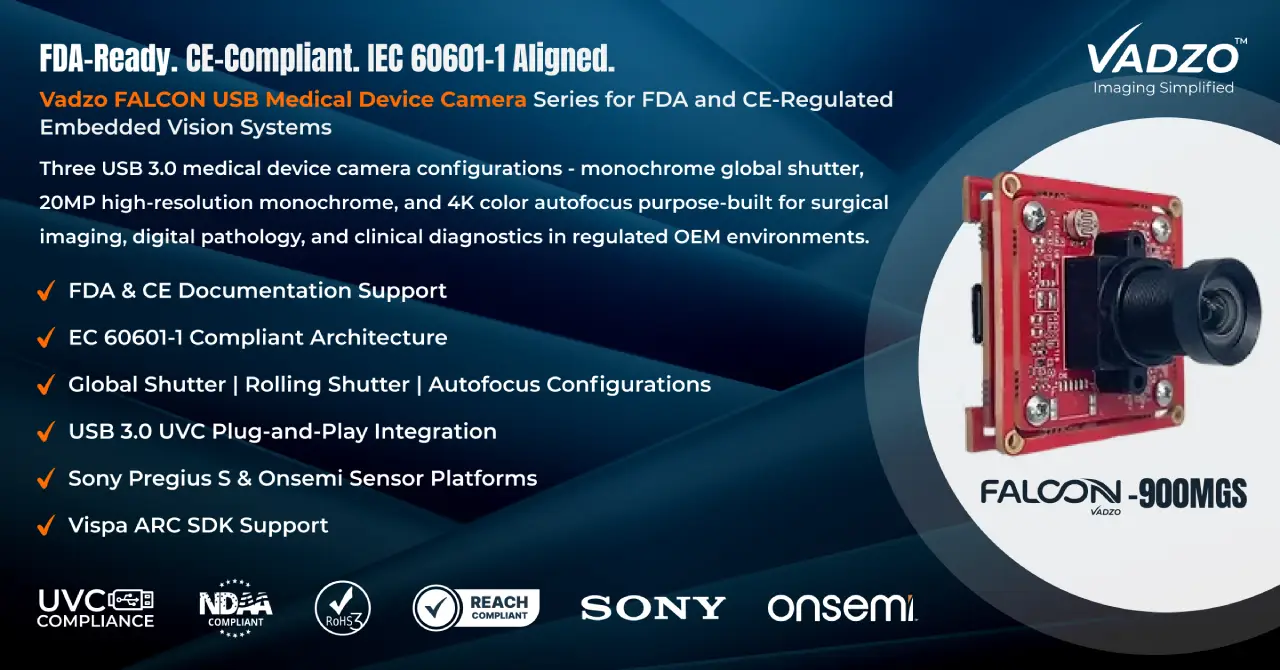
VElectrical safety class, EMC characterization, sensor architecture, and OEM compliance documentation are the variables that determine whether a medical device camera integration clears FDA 510(k) and CE regulatory submission. Vadzo Imaging's FALCON USB camera series addresses all of them across three USB 3.0 configurations purpose-built for surgical imaging, digital pathology, and clinical diagnostics.
FORT WORTH, TX / ACCESS Newswire / May 20, 2026 / Vadzo Imaging, a provider of embedded vision camera, is addressing a challenge that surfaces early in nearly every medical OEM design cycle: What does compliant hardware selection actually require when a medical device camera must survive regulatory review under FDA 21 CFR Part 820, IEC 60601-1, or CE marking directives? OEM developers building diagnostic vision systems, surgical imaging platforms, and patient monitoring instruments consistently encounter the same gap sensor specifications that alone do not determine whether a medical device camera is suitable for regulated environments. Electrical safety class, EMC performance, biocompatibility of materials in patient-accessible zones, and a camera manufacturer's ability to supply documentation for technical files all determine whether a medical device camera integration clears regulatory submission or stalls there.
Vadzo's response is its FALCON USB camera series, three USB 3.0 medical device camera products built on Sony and Onsemi sensor platforms that OEM developers and system integrators are actively qualifying for digital pathology camera systems, microscopy imaging camera platforms, surgical vision system deployments, and clinical imaging camera applications. Across all three, Vadzo delivers OEM medical camera configurations matched precisely to the imaging resolution, dynamic range, and interface requirements that medical device vision system design demands and backs them with documentation and customization services that regulated product development requires.
Why USB 3.0 is the Interface Medical OEM Developers Select First
Medical device integration environments impose constraints that GigE and MIPI configurations do not always satisfy cleanly. Board space in handheld diagnostic instruments and modular surgical consoles is limited. Host compute platforms in embedded medical systems run Linux or Windows acquisition stacks, where USB 3.0 provides immediate enumeration without custom driver stacks. USB 3.0 delivers up to 5 Gbps of sustained bandwidth, sufficient for uncompressed RAW streaming from sensors up to 20MP at clinically useful frame rates, over cable runs practical within a medical cart, surgical tower, or portable diagnostic enclosure.
For medical OEM integration, USB also simplifies the isolation architecture. Class II USB-powered peripherals avoid the leakage current concerns that introduce complexity into IEC 60601-1 compliance analysis when line-powered imaging hardware is placed in patient-accessible locations. This makes USB 3.0 the pragmatic starting point for most embedded medical imaging and healthcare imaging camera deployments where the host system, not the camera, carries the primary electrical safety classification. This USB-powered configuration is the baseline starting point for any IEC 60601 camera selection where patient-accessible placement and minimal leakage current contribution are both non-negotiable.
"Engineering teams qualifying a medical device camera for FDA or CE submission ask us the same core questions: What is the electrical safety class, what EMC characterization data exists, and can you support the technical file? The FALCON series is built to answer all three, starting with the right sensor, on the right interface, with the right OEM documentation support behind it," says Alwin Vincent, Product Manager, Vadzo Imaging.
FALCON-900MGS: 3MP IMX900 Monochrome Global Shutter USB 3.0 Camera
Fluorescence microscopy, UV-excited pathology imaging, and IR-based clinical applications share a hard requirement: no color filter array, no rolling shutter distortion, and maximum photon efficiency across the visible and near-infrared spectrum. The FALCON 900MGS (IMX900 USB 3.0 Camera) is Vadzo's monochrome global shutter medical device camera built around the Sony Pregius S IMX900 CMOS sensor, a stacked BSI CMOS device with 2.25 µm pixel pitch delivering 3MP (2064×1552) with simultaneous all-pixel exposure. Global shutter eliminates the spatial-temporal artifact that rolling shutter introduces when imaging moving tissue samples on motorized stages, pulsed fluorescence illumination systems, or strobe-synchronized diagnostic imaging camera setups. The IMX900 BSI architecture improves NIR quantum efficiency directly, making this IMX900 Monochrome Camera the correct embedded vision medical camera choice wherever wavelength-specific sensitivity determines clinical data quality.
Key specs: 3MP (2064×1552) | Sony Pregius S IMX900 | Global Shutter | 1/3.1" 2.25µm BSI Pixel | USB 3.0 | Quad HDR (up to 120dB) | S-Mount (M12 Standard) | -30⁰C to 70⁰C
FALCON-2020MRS: 20MP AR2020 Monochrome USB 3.0 Camera
Digital pathology and high-magnification clinical microscopy require spatial resolution that 3MP and 5MP sensors simply cannot provide. Whole-slide imaging instruments, dermatoscopy platforms, and high-content screening systems need sensors resolving features at the submicron scale across wide fields without stitching artifacts. The FALCON 2020MRS delivers 20MP (5120×3840) through the Onsemi Hyperlux LP AR2020 Imaging Sensor, a monochrome platform with 1.4 µm pixel pitch on a 1/1.8" optical format that maximizes compatibility with standard C-mount microscopy optics. As a medical device camera for digital pathology, a single frame from this AR2020 Monochrome USB 3.0 Camera captures field detail that lower-resolution sensors approximate only through multi-acquisition stitching workflows that introduce motion-induced registration errors and extend acquisition time in regulated instrument pipelines. The 4K AR2020 Rolling Shutter USB 3.0 Camera streams over USB 3.0 with no frame grabber hardware, integrating directly into the acquisition pipelines that medical device software stacks already manage.
Key specs: 20MP (5120×3840) | Onsemi Hyperlux LP AR2020 | Rolling Shutter | 1/1.8" 1.4µm Pixel | USB 3.0 | HDR | S-Mount (M12 Standard) | -30⁰C to 70⁰C
FALCON-1335CRA: 4K AR1335 Color Autofocus USB 3.0 Camera
Surgical imaging, endoscopy integration, and patient monitoring applications demand color fidelity, sufficient resolution to render tissue texture and vascular structure accurately, and autofocus performance that tracks depth changes in real time without operator intervention. The FALCON 1335CRA is Vadzo's medical device camera for color clinical imaging, built on the Onsemi AR1335 at 13MP (4208×3120) with an integrated autofocus mechanism that resolves depth across a surgical field or close-up clinical scenario without manual focus adjustment. At 4K, the FALCON 1335CRA delivers pixel density that surgical vision system developers need to maintain detail in zoomed-in ROI views and supply AI inference pipelines with high-resolution input for tissue classification, lesion detection, and instrument tracking. The 1080p AR1335 Color USB Camera output mode enables real-time streaming at lower resolution for latency-sensitive display, while retaining 4K AR1335 USB 3.0 Camera capture capability for frame-by-frame clinical review, making this AR1335 Color Autofocus USB 3.0 Camera a practical single-device solution for both surgical display and diagnostic review within one integrated platform.
Key specs: 13MP (4208×3120) | Onsemi AR1335 | Rolling Shutter | 1/3.2" 1.1µm Pixel | USB 3.0 | VCM Based Autofocus with Focus Range of 100mm to Infinity | -30⁰C to 70⁰C
Vispa ARC SDK: Camera Control for Medical Device Software Integration
All three FALCON USB camera products are supported by Vadzo's Vispa ARC SDK, providing API-level control over image acquisition parameters, exposure, gain, white balance, region of interest configuration, and firmware updates from the host application. Vispa ARC supports C, C++, and Python with compatibility across Windows and Linux, aligning with the development environments that medical device software teams operate in under IEC 62304 software lifecycle processes. The SDK delivers direct camera control without requiring OEM developers to manage low-level USB descriptor handling or custom kernel driver development, reducing integration time and the software validation scope that regulated medical device software submissions require.
Medical Device Vision System Applications
The FALCON USB camera series covers the full scope of medical device imaging modalities that medical OEM development teams encounter. Each configuration maps directly to the imaging environment it is designed for.
Digital Pathology and Microscopy Imaging: The FALCON 2020MRS 20MP AR2020 Monochrome USB 3.0 Camera serves whole-slide imaging instruments, high-content analysis platforms, and clinical microscopy camera applications where spatial resolution determines diagnostic data integrity. The FALCON 900MGS 3MP IMX900 Mono USB 3.0 Camera handles fluorescence and NIR-band microscopy imaging camera deployments where monochrome global shutter sensitivity is non-negotiable and pulsed illumination synchronization is a core acquisition requirement.
Surgical Imaging and Endoscopy: The FALCON 1335CRA AR1335 Color USB 3.0 Camera delivers color fidelity, resolution, and autofocus for surgical imaging camera and endoscopy-adjacent embedded systems where tissue visibility and real-time depth tracking define procedural outcome quality. Its autofocus mechanism operates continuously across depth changes without host-side intervention, fitting directly into surgical vision system architectures where hands-free imaging is a design requirement.
Patient Monitoring and Clinical Diagnostics: The FALCON 1335CRA AR1335 USB 3.0 Camera and FALCON 900MGS 3MP IMX900 USB Camera both address patient monitoring camera and diagnostic vision system deployments where image consistency, USB integration simplicity, and sensor-level performance need to coexist in a single validated hardware configuration. Both function as capable healthcare imaging camera across the clinical environments where these attributes matter simultaneously.
Digital Pathology and Whole-Slide Microscopy Imaging: Whole-slide imaging at 20× and 40× magnification requires pixel density sufficient to resolve nuclear morphology, chromatin structure, and cellular boundaries across a wide field in a single acquisition, a requirement that sensors below 12MP cannot meet without multi-frame stitching workflows that introduce registration errors and extend scan time. The FALCON-2020MRS 20MP monochrome output captures sufficient field area per frame at standard C-mount microscopy magnifications to reduce or eliminate stitching entirely. Rolling shutter is the correct architecture for bright-field continuous illumination pathology workflows where pulsed excitation is not involved. HDR handles the contrast range between stained tissue regions and unstained background without clipping either end. USB 3.0 integrates directly into acquisition pipelines without a frame grabber, reducing instrument BOM and software validation scope.
Fluorescence and NIR Microscopy Imaging: Fluorescence microscopy and NIR-band clinical imaging require a sensor with no color filter array absorbing the excitation wavelength, simultaneous all-pixel capture to prevent spatial-temporal distortion under pulsed illumination, and BSI pixel architecture for maximum photon efficiency at low signal levels. The FALCON-900MGS global shutter eliminates the frame distortion that rolling shutter introduces when imaging under pulsed fluorescence excitation or strobe-synchronized illumination systems. Monochrome capture preserves the full NIR signal without color filter absorption losses. BSI architecture improves quantum efficiency directly at the wavelengths fluorescence and NIR clinical applications depend on. The Sony Pregius S IMX900 Quad HDR mode extends dynamic range to 120 dB for high-contrast fluorescence scenes where dim marker signal and bright background coexist in the same frame.
Surgical Imaging and Endoscopy-Adjacent Systems: Surgical vision systems and endoscopy-adjacent platforms require accurate color rendering for tissue and vascular structure, resolution sufficient to maintain detail in zoomed ROI views, and autofocus that tracks depth changes across a surgical field without operator intervention. The FALCON-1335CRA VCM-based autofocus resolves depth continuously from 100mm to infinity without host-side intervention, fitting directly into surgical vision system architectures where hands-free imaging is a design requirement. Factory-calibrated color correction matrix delivers accurate tissue color rendering under surgical illumination without per-unit calibration on the integrator side. At 4K, the 13MP AR1335 sensor supplies AI inference pipelines with sufficient resolution for tissue classification, lesion detection, and instrument tracking. The 1080p output mode enables real-time surgical display at lower latency while retaining 4K capture capability for frame-by-frame clinical review within one integrated platform.
Patient Monitoring and Clinical Diagnostics: Patient monitoring and clinical diagnostic instruments require consistent image output across variable clinical illuminants, USB integration that enumerates driver-free on Windows and Linux acquisition stacks, and sensor configurations validated for the specific imaging modality, color for patient-facing monitoring, monochrome for wavelength-specific diagnostic imaging. The FALCON-1335CRA color output with factory-calibrated white balance delivers consistent skin tone and tissue color rendering across the mixed fluorescent, LED, and daylight illuminants of clinical environments without per-installation tuning. The FALCON-900MGS handles diagnostic modalities requiring NIR sensitivity or pulsed illumination synchronization where color information has no clinical value and photon efficiency determines diagnostic data quality. Both cameras enumerate driver-free as standard USB 3.0 UVC devices on Windows and Linux, reducing software integration scope and the validation effort regulated medical device software submissions require under IEC 62304.
What the FALCON USB Camera Series Shares: Vadzo's OEM Commitment to Medical Integrators
Across all three FALCON medical device camera products, Vadzo provides OEM services that medical device development teams depend on beyond camera hardware alone. Full OEM camera customization covers board-level redesigns, firmware modifications, medical optics integration for C-mount and custom lens configurations, and thermal management adaptation for sealed enclosure designs. Documentation packages supporting IEC 60601-1 electrical safety analysis, CE marking technical files, and FDA 510(k) design history file compilation are available on request. Vadzo's applications engineering team engages directly with OEM development teams on medical imaging compliance documentation scope from early design-in through regulatory submission. ISP tuning is calibrated against the clinical imaging environments each sensor configuration targets. Volume pricing, production support, and direct applications engineering engagement for design-in and regulatory review assistance are available to qualified OEM development teams.
Frequently Asked Questions (FAQs)
1) What interface is best for a medical device camera used in IEC 60601-1 regulated environments?
USB 3.0 is the interface most OEM medical device developers select first, and for good reason. A USB-powered camera operates as a Class II peripheral with no mains-connected current path, which means it contributes negligibly to the patient leakage current budget defined under IEC 60601-1. Line-powered GigE cameras in patient-accessible zones require significantly more complex isolation analysis, adding scope to both the safety assessment and the technical file.
USB 3.0 also delivers up to 5 Gbps of sustained bandwidth sufficient for uncompressed RAW streaming from sensors up to 20MP at clinically relevant frame rates over cable lengths practical within a medical cart, surgical tower, or portable diagnostic enclosure. Vadzo's FALCON USB camera series is built specifically on this interface rationale, offering three medical device camera configurations across global shutter, high-resolution monochrome, and 4K color autofocus platforms, each backed by IEC 60601-1 electrical safety documentation support for OEM technical file compilation.
2) What resolution does a medical device camera need for digital pathology scanning without image stitching?
Whole-slide imaging at standard 20× and 40× magnification requires enough pixel density to resolve nuclear morphology, chromatin structure, and cellular boundary detail across a wide field in a single acquisition. Sensors below 12MP typically cannot cover enough field area at these magnifications without requiring multi-frame stitching, a process that introduces motion-induced registration errors, extends scan time, and adds computational overhead to the instrument pipeline.
At 20MP, a medical device camera built on the Onsemi Hyperlux LP AR2020 sensor captures sufficient field area per frame at standard C-mount microscopy magnifications to reduce or eliminate stitching entirely. The 1.84 µm pixel pitch resolves subcellular features that coarser pixel architectures miss at equivalent magnification. For OEM developers building digital pathology instruments, this means fewer acquisition passes, cleaner slide data, and a simpler instrument software architecture, all of which reduce both development complexity and the software validation scope required under IEC 62304.
3) How do I choose between a global shutter and a rolling shutter for a clinical imaging application?
The decision maps directly to your illumination architecture and whether your subject moves during the exposure window. Global shutter captures all pixels simultaneously, making it essential for pulsed fluorescence excitation, strobe-synchronized clinical setups, and motorized stage microscopy. In these configurations, rolling shutter introduces a spatial-temporal distortion across the frame, where different rows are captured at different moments, which invalidates fluorescence intensity data and creates measurement error in quantitative imaging applications.
Rolling shutter sensors are the correct choice for bright-field pathology, surgical color imaging, and continuous-illumination clinical inspection, where pulsed illumination is not involved and motion within a single frame is not a variable. Vadzo's FALCON USB medical device camera series covers both scenarios. The FALCON 900MGS 3MP IMX900 Monochrome Global Shutter USB 3.0 Camera delivers global shutter performance on the Sony Pregius S IMX900 for pulsed and NIR clinical imaging, while the FALCON 2020MRS 20MP AR2020 Monochrome USB 3.0 Camera and FALCON 1335CRA 13MP AR1335 Color Autofocus USB 3.0 Camera provide rolling shutter configurations for pathology and surgical applications, respectively.
4) What documentation should a medical device camera vendor provide to support an FDA 510(k) or CE marking technical file?
At minimum, a camera vendor supporting a regulated medical device integration should supply electrical safety characterization data relevant to IEC 60601-1, EMC emissions and immunity test reports, a declaration of conformity, and materials declarations covering RoHS and REACH compliance. For design history file purposes under FDA 21 CFR Part 820, component-level datasheets, interface specifications, and firmware version control documentation are equally important.
For CE marking under EU MDR 2017/745, the harmonized standard set typically includes IEC 60601-1 for electrical safety, IEC 60601-1-2 for EMC, and ISO 14971 inputs for risk management. The gap most OEM teams encounter is that consumer-grade camera vendors have none of this documentation on file, which stalls the submission at the technical file review stage. Vadzo provides a documentation package for the FALCON USB medical device camera series covering these inputs for qualified OEM development teams. Contact the applications engineering team to scope the deliverables against your specific submission requirements.
5) Which SDK supports IEC 62304 compliant software development for an embedded medical imaging system?
IEC 62304 requires software lifecycle documentation that covers third-party components integrated into the medical device software stack, including version traceability, change control records, and test evidence for the functions your application depends on. Most camera SDKs from consumer-grade vendors are undocumented at this level, which forces medical device software teams to generate their own validation evidence from scratch, adding weeks to the software validation plan.
Vadzo's Vispa ARC SDK supports the FALCON USB medical device camera series with version-tracked firmware releases, documented API change logs, and release notes that provide the traceability inputs IEC 62304 Class B and Class C software lifecycle documentation requires. The SDK supports C, C++, and Python across Windows and Linux, covering the development environments most embedded medical imaging teams already operate in. This reduces the third-party component integration scope in your software validation plan and shortens the path from prototype to regulatory-ready software build.
Availability
The FALCON 900MGS IMX900 Monochrome USB 3.0 Camera, FALCON 2020MRS 20MP AR2020 Monochrome USB 3.0 Camera, and FALCON 1335CRA 4K AR1335 Color USB 3.0 Camera are available for OEM evaluation. Evaluation units, technical documentation, Vispa ARC SDK access, and integration support are available directly from Vadzo Imaging at www.vadzoimaging.com. Volume pricing, firmware customization, medical optics integration, and enclosure design services are available upon request. For inquiries, contact the Vadzo sales team at [email protected].
About Vadzo Imaging
Vadzo Imaging develops high-performance embedded and machine vision cameras for OEMs and system integrators building next-generation intelligent systems. The company delivers imaging platforms across USB, MIPI, GigE, Wi-Fi, and SerDes interfaces, supporting applications in industrial automation, robotics, smart surveillance, smart city infrastructure, edge AI, and regulated healthcare through its medical device camera portfolio. Beyond hardware, Vadzo provides end-to-end imaging expertise, including sensor integration, ISP tuning, firmware development, and OEM camera customization services that accelerate development and deployment at scale.
Media Contact
Alwin Vincent
Vadzo Imaging
Email: [email protected]
LinkedIn: Vadzo Imaging
YouTube: Vadzo Imaging
X: Vadzo Imaging
SOURCE: Vadzo Imaging